Beranda
/ Electron Microscope Diagram Of Animal Cell - Electron Microscopic Study Of Cell And Organelles Important - The specimen is most often an ultrathin section less than 100 nm thick or a suspension on a grid.
Electron Microscope Diagram Of Animal Cell - Electron Microscopic Study Of Cell And Organelles Important - The specimen is most often an ultrathin section less than 100 nm thick or a suspension on a grid.
Electron Microscope Diagram Of Animal Cell - Electron Microscopic Study Of Cell And Organelles Important - The specimen is most often an ultrathin section less than 100 nm thick or a suspension on a grid.. Image viewing, development and recording techniques 6. It's made up of phospholipids and also contains protein and carbohydrate molecules. Feb 15, 2020 · animal cell size and shape. High voltage modern electron … They have an outer cell wall that gives them shape.
The specimen is most often an ultrathin section less than 100 nm thick or a suspension on a grid. As is the case with animal cells, the cell membrane in plants is a lipid bilayer. The microscope parts work together in hospitals and in forensic labs, for scientists and students, bacteriologists and biologists so that they may view bacteria, plant and animal cells and tissues, and various microorganisms the world over. Animal cells come in all kinds of shapes and sizes, with their size ranging from a few millimeters to micrometers. Furthermore, it is easy to distinguish between a plant and animal cell diagram just by inspecting the presence or absence of a cell wall.
Q14 Draw A Large Diagram Of An Animal Cell As Seen Through An Electron Microscope Label The Parts That Science Tissues 11500353 Meritnation Com from s3mn.mnimgs.com Animal cells come in all kinds of shapes and sizes, with their size ranging from a few millimeters to micrometers. Preparing a wet mount of a specimen is the technique typically used to view plant and animal cells using a microscope.this page provides step by step instructions on slide preparation as well as videos at the bottom of page. High voltage modern electron … Feb 15, 2020 · animal cell size and shape. It's made up of phospholipids and also contains protein and carbohydrate molecules. Transmission electron microscopy (tem) is a microscopy technique in which a beam of electrons is transmitted through a specimen to form an image. The specimen is most often an ultrathin section less than 100 nm thick or a suspension on a grid. As is the case with animal cells, the cell membrane in plants is a lipid bilayer.
High voltage modern electron …
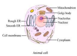
Animal cells come in all kinds of shapes and sizes, with their size ranging from a few millimeters to micrometers. It's made up of phospholipids and also contains protein and carbohydrate molecules. Preparing a wet mount of a specimen is the technique typically used to view plant and animal cells using a microscope.this page provides step by step instructions on slide preparation as well as videos at the bottom of page. The nuclear membrane has pores. They have an outer cell wall that gives them shape. Furthermore, it is easy to distinguish between a plant and animal cell diagram just by inspecting the presence or absence of a cell wall. Here, the cell membrane is involved in a number of functions including containing cell organelles, transportation of molecules in and out of the cell as well as cell communication. As is the case with animal cells, the cell membrane in plants is a lipid bilayer. Feb 15, 2020 · animal cell size and shape. Principle of electron microscope 2. Though this animal cell diagram is not representative of any one particular type of cell, it provides insight into the primary organelles and the intricate internal structure of most animal cells. Just under the rigid cell wall is the more fluid cell membrane. Use of electron microscope 7.
Preparing a wet mount of a specimen is the technique typically used to view plant and animal cells using a microscope.this page provides step by step instructions on slide preparation as well as videos at the bottom of page. Use the following animation to explore bacterial structure. Image viewing, development and recording techniques 6. As is the case with animal cells, the cell membrane in plants is a lipid bilayer. Just under the rigid cell wall is the more fluid cell membrane.
Draw A Large Diagram Of An Animal Cell As Seen Through An Electron Microscope Label The Parts That Brainly In from hi-static.z-dn.net The cytoplasm enclosed within the cell membrane does not exhibit much structure when viewed by electron microscopy. Even though plant and animal cells are eukaryotic and share a few cell organelles, plant cells are quite distinct when compared to animal cells as they perform different functions. Just under the rigid cell wall is the more fluid cell membrane. At certain points, the nuclear membrane is continuous with the endoplasmic reticulum. High voltage modern electron … As is the case with animal cells, the cell membrane in plants is a lipid bilayer. Components of electron microscope 4. Feb 15, 2020 · animal cell size and shape.
Some of these differences can be clearly understood when the cells are examined under an electron microscope.
High voltage modern electron … Preparing a wet mount of a specimen is the technique typically used to view plant and animal cells using a microscope.this page provides step by step instructions on slide preparation as well as videos at the bottom of page. They have an outer cell wall that gives them shape. Just under the rigid cell wall is the more fluid cell membrane. Components of electron microscope 4. The nuclear membrane has pores. Here, the cell membrane is involved in a number of functions including containing cell organelles, transportation of molecules in and out of the cell as well as cell communication. As is the case with animal cells, the cell membrane in plants is a lipid bilayer. Use the following animation to explore bacterial structure. Furthermore, it is easy to distinguish between a plant and animal cell diagram just by inspecting the presence or absence of a cell wall. Under the electron microscope, the nuclear membrane is seen to consist of inner and outer layers of electron dense material and the middle one of electron transparent substance. Feb 15, 2020 · animal cell size and shape. It's made up of phospholipids and also contains protein and carbohydrate molecules.
Here, the cell membrane is involved in a number of functions including containing cell organelles, transportation of molecules in and out of the cell as well as cell communication. Principle of electron microscope 2. Transmission electron microscope (tem) 3. Transmission electron microscopy (tem) is a microscopy technique in which a beam of electrons is transmitted through a specimen to form an image. Furthermore, it is easy to distinguish between a plant and animal cell diagram just by inspecting the presence or absence of a cell wall.
Animal Cell Definition Structure Parts Functions And Diagram from microbenotes.com The microscope parts work together in hospitals and in forensic labs, for scientists and students, bacteriologists and biologists so that they may view bacteria, plant and animal cells and tissues, and various microorganisms the world over. High voltage modern electron … The nuclear membrane has pores. Though this animal cell diagram is not representative of any one particular type of cell, it provides insight into the primary organelles and the intricate internal structure of most animal cells. Furthermore, it is easy to distinguish between a plant and animal cell diagram just by inspecting the presence or absence of a cell wall. Preparing a wet mount of a specimen is the technique typically used to view plant and animal cells using a microscope.this page provides step by step instructions on slide preparation as well as videos at the bottom of page. Some of these differences can be clearly understood when the cells are examined under an electron microscope. Transmission electron microscopy (tem) is a microscopy technique in which a beam of electrons is transmitted through a specimen to form an image.
At certain points, the nuclear membrane is continuous with the endoplasmic reticulum.
Just under the rigid cell wall is the more fluid cell membrane. The cytoplasm enclosed within the cell membrane does not exhibit much structure when viewed by electron microscopy. Under the electron microscope, the nuclear membrane is seen to consist of inner and outer layers of electron dense material and the middle one of electron transparent substance. Here, the cell membrane is involved in a number of functions including containing cell organelles, transportation of molecules in and out of the cell as well as cell communication. After reading this article you will learn about: They have an outer cell wall that gives them shape. Components of electron microscope 4. Use the following animation to explore bacterial structure. High voltage modern electron … Even though plant and animal cells are eukaryotic and share a few cell organelles, plant cells are quite distinct when compared to animal cells as they perform different functions. The specimen is most often an ultrathin section less than 100 nm thick or a suspension on a grid. The microscope parts work together in hospitals and in forensic labs, for scientists and students, bacteriologists and biologists so that they may view bacteria, plant and animal cells and tissues, and various microorganisms the world over. Image viewing, development and recording techniques 6.
Berbagi :
Posting Komentar
untuk "Electron Microscope Diagram Of Animal Cell - Electron Microscopic Study Of Cell And Organelles Important - The specimen is most often an ultrathin section less than 100 nm thick or a suspension on a grid."


Posting Komentar untuk "Electron Microscope Diagram Of Animal Cell - Electron Microscopic Study Of Cell And Organelles Important - The specimen is most often an ultrathin section less than 100 nm thick or a suspension on a grid."